馬特里病
1. 疾病描述
1.病原概述
病原型別:原生生物(protest)
病原環境:海水
學名:Mikrocytos mackini
病名(及俗名):Denman Island disease 或稱 Microcell disease of Pacific oyster.
最早發現者:
OIE狀況:表列(Listed)
病原摘要:
Mikrocytos mackini 為一原生動物大小約2-4um,目前所知尚無其它strains (Farley 1988),目前對於此病原於離開宿主後存活情形不祥,病原可經水而水平傳染。
人畜共通:No
病原分類
2.病原分類
目前已知Mikrocytos mackini 與其它 microcells 屬如 Bonamia 等無關( 如 B. ostreae, B. exitiosa, B. roughleyi and other Bonamia spp.) (Carnegie and Cochennec-Laureau 2004).
疾病特性:
疾病型態及流行病學:急性或慢性型態、器官傳播分佈情形
本病主要流行於北美西岸如加拿大西岸、美國華盛頓州等地區,當水溫低於10℃ 維持3-4個月後其發生率與致死率則常可達族群一半,顯然約有一半是具有抵抗性,Hervio 1996 研究指出要引發牡蠣感染本病須於10℃以下連續3月以上才會成立,本疾病之傳播不須任何煤介。
臨床症狀及病理學:
臨床上牡蠣通常於春天時後發生死亡,並可於其軟組織中發現直徑約5 mm 大小之黃綠色(yellow green)膿皰(pustules),其殼上亦有棕色之疤(scar),但發現感染死亡之牡蠣其它狀況仍良好(good condition),但這些並非本病之特異病變。
病理學上病材可固定於Davidson’s solution 或中性福馬林(10% buffered formalin)。
病原(Microcells) 可於小囊結締組織細胞之細胞質(within the cytoplasm of cesicular connective tissue cells) 中發現,特別於膿皰(pustules)周圍之結締組織,同時亦有血細胞之浸潤 (haemocyte infiltration ) 及壞死之細胞 (Bower 2007)
Mikrocytos mackini 直徑約2-4Mm 在病灶區除上述組織外亦會感染肌細胞(myocytes )及血球細胞(haemocytes)。
病原致病性意義:實際魚場及田間發病、抗藥性等情形
Mikrocytos mackini 可感染多種oysters(牡蠣)如 Crassostrea
gigas (PaciWc Oysters) 、Crassostrea virginica (eastern oysters) 、Ostrea edulis (European Xat oysters) 及 Ostrea conchaphila (Olympia oysters)等(Bower et al 2005),但有些 clam 如 Panope abrupta 則具有抵抗性。
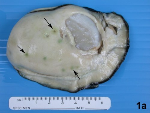
Crassostrea gigas 由殼移出於箭所指可明顯看出 Mikrocytos mackini 充滿於 vesicular connective tissue cells 肉眼上呈現徑約5mm 大小之黃綠色(yellow green)膿皰(pustules)。(Bower 2007)
2. 診斷方式
組織壓片:以乾淨之刀片切病灶區(pustule) 直接壓至乾淨之玻片後並以Wright-Giemsa染色後以1000x 之油鏡直接觀查病原---此方法之特異性不高。
組織病理切片
分子生物學-PCR(Carnegie et al 2003)
選擇病牡蠣或剛死亡之檢體
DNA之抽取:可使用一般之kit 如 (e.g. DNeasy Kit; QIAGEN).
引子對分別為MIKROCYTOS-F (AGATGGTTAATGAGCCTCC) 及 MIKROCYTOS-R (GCGAGGTGCCACAAGGC).其所使用之反應條件分別為
94℃ 10分鐘,之後40 個循環分別為94℃ 1分、60.5℃ 分、72℃ 1分,最後72℃ 10 分。其產物大小為546bp。
in-situ hybridisation (ISH).原位雜交法:選擇膿皰(pustules)周圍組織 及內收肌(adductor mucles) 方法可參考Meyer 2005。
3. 治療方法
4. 預防措施
目前無疫苗、化學治療劑及免疫刺進劑可供使用(vaccination: none、 chemotherapy: none、immunostimulation : none)亦無發展出有效之抗病品種。因此可先行採收3年內(最好於第3年2月前)已達上市大小之牡蠣、在6月前不要與可能發病之牡蠣一起養殖。
5. 參考資料
Bower SM, Bate K, Meyer GR. Susceptibility of juvenile Crassostrea gigas and resistance of Panope abrupta to Mikrocytos mackini. J. Invertebr. Pathol., 88: 95-99, 2005.
Bower SM, Carnegie RB, Goh B, Jones SRM, Lowe G.J, Mak MWS. Preferential PCR amplification of parasitic protistan small subunit rDNA from metazoan tissues. J. Eukaryotic Microbiol., 51: 325-332, 2004.
Bower SM. Synopsis of Infectious Diseases and Parasites of Commercially Exploited Shellfish: Mikrocytos mackini (Denman Island Disease) of Oysters. 2007.
Carnegie RB, Cochennec-Laureau N. Microcell parasites of oysters: recent insights and future trends. Aquatic Living Resources 17: 519-528,
2004.
Carnegie RB, Meyer G.R, Blackbourn J, Cochennec-Laureau N, Berthe FCJ, Bower SM. Molecular detection of the oyster parasite Mikrocytos mackini and a preliminary phylogenetic analysis. Dis. Aquat. Org., 54: 219-227, 2003.
Farley CA, Wolf PH, Elston RA. A long-term study of 'microcell' disease with a description of a new genus, Mikrocytos (g. n.), and two new species, Mikrocytos mackini (sp. n.) and Mikrocytos roughleyi (sp. n.). Fishery Bull., 86: 581-593,1988.
Hervio D, Bower SM, Meyer GR. Detection, isolation and experimental transmission of Mikrocytos mackini, a microcell parasite of PaciWc oysters Crassostrea gigas (Thunberg). J. Invertebr. Pathol. 67: 72–79,1996.
Meyer GR, Bower SM, Carnegie RB. Sensitivity of a digoxigenin-labelled DNA probe in detecting Mikrocytos mackini, causative agent of Denman Island disease (mikrocytosis) in oysters. J. Invertebr. Pathol. 88: 89-94, 2005.
相關連結: